Description
Overview
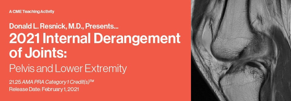
Internal Derangement of Joints 2021: Pelvis and Lower Extremity (CME VIDEOS) is a comprehensive continuing education activity focused on MR imaging assessment of musculoskeletal disorders affecting the pelvis, hip, knee, ankle, and foot. This course emphasizes essential anatomy, physiology, and pathology to enhance the interpretation of imaging findings in lower extremity joints, comparing MR imaging to other diagnostic methods. On-demand radiology education — accessible anytime, anywhere.
Agenda
This CME video series provides detailed presentations on key topics related to internal derangements of joints in the pelvis and lower extremity. Each session is designed to strengthen expertise in MR image assessment and clinical interpretation.
- Articular Disorders of Synovium-Lined Joints: Joint Morphology and General Abnormalities, Specific Inflammatory and Degenerative Disorders, Tumors and Tumor-Like Disorders — Donald L. Resnick, M.D.
- Focus Session: Osteonecrosis Versus Insufficiency Fractures: Emphasis on the Hip and Knee — Donald L. Resnick, M.D.
- Chondral, Osteochondral and Subchondral Injuries: Anatomy, Pathophysiology, Terminology and MR Imaging — Donald L. Resnick, M.D.
- Cartilage Imaging: Routine and Advanced Methods — Christine B. Chung, M.D.
- Stress-Related Abnormalities of the Skeleton with Emphasis on the Pelvis and Lower Extremity — Mini N. Pathria, M.D.
- Focus Session: Muscle Disorders: Anatomy Pathophysiology and General Abnormalities — Mini N. Pathria, M.D.
- Muscles and Tendons About the Pelvis and Hip: Anatomy, Strains and Tears — Mini N. Pathria, M.D.
- Labral Abnormalities and External and Internal Femoroacetabular Impingement — Donald L. Resnick, M.D.
- Focus Session: Important Entrapment Neuropathies of the Pelvis and Lower Extremity — Evelyne A. Fliszar, M.D.
- Meniscus: Structure, Function and Patterns of Failure — Donald L. Resnick, M.D.
- Discoid Menisci and Other Anomalies — Donald L. Resnick, M.D.
- Focus Session: Bone Marrow: Normal and Abnormal with Emphasis on MRI — Evelyne A. Fliszar, M.D.
- Anatomy, Biomechanics and Footprints of Injury — Donald L. Resnick, M.D.
- Anterior Cruciate Ligament — Brady Huang, M.D.
- Posterior Cruciate Ligament — Brady Huang, M.D.
- Medial Supporting Structures of the Knee — Donald L. Resnick, M.D.
- Lateral Supporting Structures of the Knee — Brady Huang, M.D.
- Postoperative Ligaments with Emphasis on the Anterior Cruciate Ligament — Brady Huang, M.D.
- Patellofemoral Maltracking and Patellar Instability/Dislocation — Lucas Hiller, M.D., M.S.E.
- Quadriceps/Patellar Tendons, Fat Pads, Bursae and Plicae — Mini N. Pathria, M.D.
- Popliteal Fossa — Mini N. Pathria, M.D.
- Focus Session: MRI/Arthroscopy Correlation — Eric Y. Chang, M.D.
- Osteomyelitis, Septic Arthritis and Soft Tissue Infection with Emphasis on the Diabetic Foot — Karen C. Chen, M.D.
- Fractures/Dislocations of the Ankle and Foot: Role of CT Scanning — Tudor Hughes, M.D., FRCR
- Tumors and Tumor-Like Lesions of the Ankle and Foot — Edward Smitaman, M.D.
- Tendons: Normal Anatomy — Donald L. Resnick, M.D.
- Adult Acquired Flatfoot Deformity — Mini N. Pathria, M.D.
- Tendons: Tendinosis, Tenosynovitis, Tendon Tears and Other Tendon Abnormalities — Donald L. Resnick, M.D.
- Rapid Fired Case Review Session: Tarsal Coalition, Osteochondritis Dissecans of the Talus, Metatarsalgia, Plantar Aponeurosis — Edward Smitaman, M.D., Karen C. Chen, M.D., Christine B. Chung, M.D.
- Ligaments: Normal Anatomy — Donald L. Resnick, M.D.
- Ligaments: Patterns of Injury — Donald L. Resnick, M.D.
Educational Objectives
Upon completion of this CME activity, participants should be able to:
- Assess MR images in patients with internal derangements of peripheral joints.
- Articulate anatomic features fundamental to accurate interpretation of MR imaging findings in these disorders.
- Formulate a reasonable differential diagnostic list and identify the most likely diagnosis.
- Understand the pathogenesis and imaging findings associated with common disorders of the pelvis and lower extremities.
Speakers
- Donald L. Resnick, M.D.
- Christine B. Chung, M.D.
- Mini N. Pathria, M.D.
- Evelyne A. Fliszar, M.D.
- Brady Huang, M.D.
- Lucas Hiller, M.D., M.S.E.
- Eric Y. Chang, M.D.
- Karen C. Chen, M.D.
- Tudor Hughes, M.D., FRCR
- Edward Smitaman, M.D.
This series was originally released with CME credit available from 1/31/2021 through 1/31/2024.
Target Audience
This educational activity is intended for practicing radiologists, orthopedic surgeons, rheumatologists, podiatrists, sports medicine physicians, and other physicians involved in musculoskeletal disorder diagnosis and management.
Why This Topic Matters
Internal Derangement of Joints 2021: Pelvis and Lower Extremity (CME VIDEOS) provides current imaging insights essential for specialists interpreting musculoskeletal conditions in the pelvis and lower extremities. Mastery of MR imaging features facilitates clearer diagnostic decision-making in radiology and orthopedic practice.
The content supports ongoing professional development within radiology and musculoskeletal medicine through MedHub Central, enhancing clinical expertise with updated knowledge relevant to patient care involving internal joint derangements.